- This event has passed.
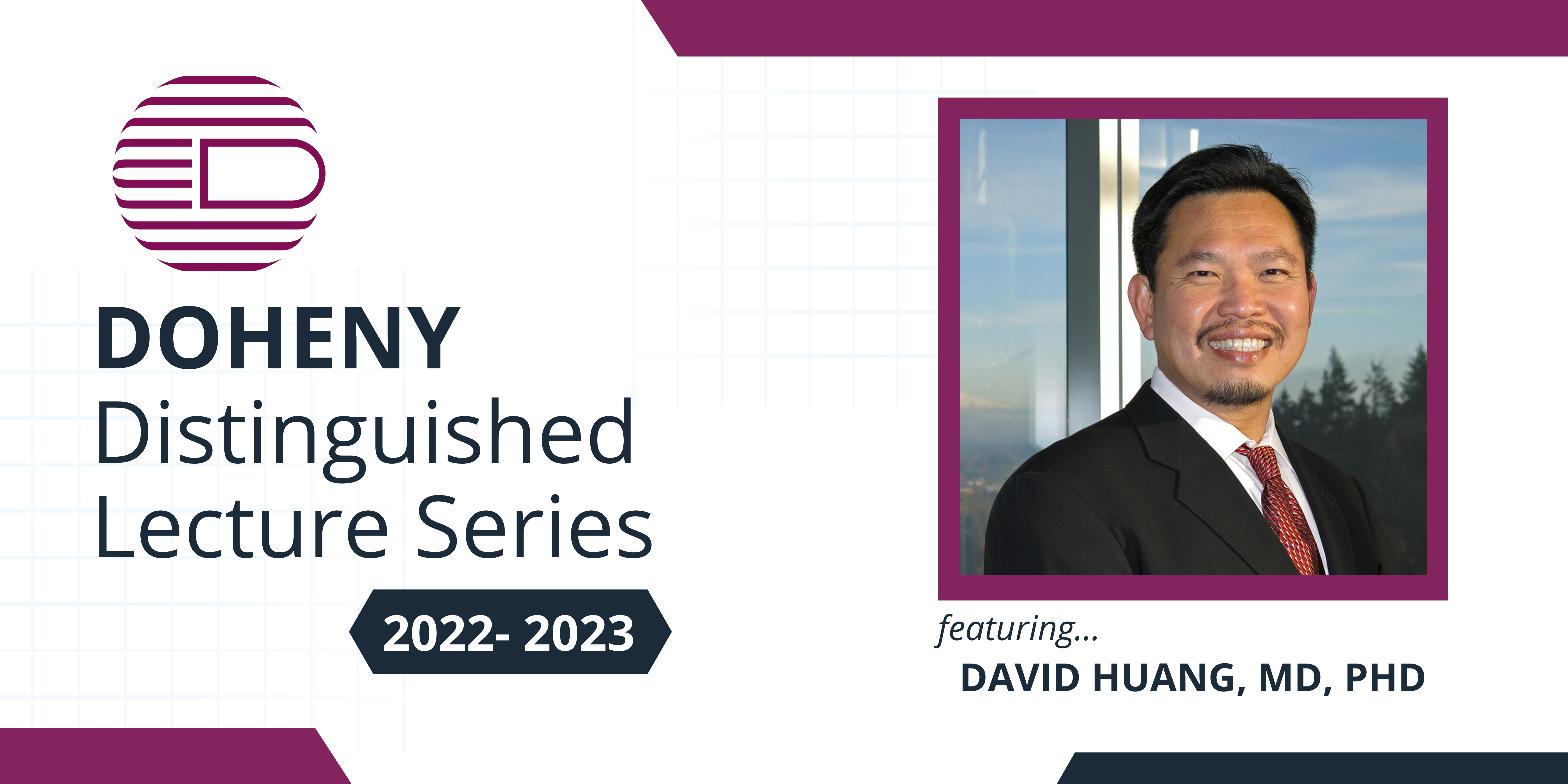
Distinguished Lecture Series – OCT Angiography and Beyond
October 7, 2022 @ 12:00 pm - 1:00 pm

Abstract
Optical coherence tomographic angiography (OCTA) is the most important new clinical OCT technology in the past decade. It became available as a clinical instrument in 2014. Since then it has been applied to a wide range of retinal and optic nerve diseases. This presentation will review the current state of the art and key clinical applications. I will also report on the development of several novel OCT technologies in our laboratory that may have important clinical applications in the near future.
One of the important recent advances in OCTA was the development of algorithms to reduce projection artifacts. Projection-resolved OCTA allows visualization of 4 distinct retinal vascular plexuses and improves the diagnosis and classification of optic nerve diseases that primarily affect the superficial plexuses, outer retinal diseases that primarily affect the deeper plexuses, and vascular diseases that affect all plexuses.
One killer app for OCTA is imaging of choroidal neovascularization (CNV). OCTA can visualize CNV below the retinal pigment epithelium better than fluorescein angiography (FA). It is now common to detect CNV that is invisible on FA and shows no retinal fluid accumulation on structural OCT. It may be important to monitor the vessel area of these nonexudative CNV, which often experience rapid geometric growth prior to the development of exudation and vision loss. In some diseases such as central serous chorioretinopathy (CSR), OCTA is useful in distinguishing fluid accumulation due to CSR versus secondary CNV.
The evaluation of diabetic retinopathy is another emerging application for OCTA. OCTA is able to quantify macular and foveal ischemia, which may serve as prognostic and pharmocodynamic marker for emerging treatments. OCTA can detect preretinal neovascularization with high sensitivity and measure their vessel area. Widefield OCTA may be useful in the early detection of proliferative diabetic retinopathy and monitoring of treatment efficacy.
Ultrawide-field OCT can image almost the entire retina or the ocular surface. Applications in retinopathy of prematurity, peripheral retinal pathologies, and corneoscleral topography will be discussed. On the other end of the spatial scale, OCT microscopy using short wavelength light (i.e. blue or green) can visualize very small structures such as bacteria and collagen lamellae. Early in vitro results will be shown. Finally, novel contrasts such as directional OCT and OCT oximetry are progressing toward clinical application. Preliminary results in human and rodents will be shown.
Bio
Dr. Huang is the Associate Director and Director of Research of Casey Eye Institute, and the Peterson Professor of Ophthalmology and Professor of Biomedical Engineering at the Oregon Health & Science University. He leads the Center for Ophthalmic Optics and Lasers (www.COOLLab.net) that conducts research on topics ranging from the development of novel instrumentation and algorithms to clinical studies in a wide variety of ophthalmic and neurological diseases. Dr. Huang is globally known for his innovations in applying laser and optical technology to eye diseases. He is a co-inventor of optical coherence tomography (OCT), a commonly used ophthalmic imaging technology with 30 million procedures performed annually worldwide. His seminal article on OCT, published in Science in 1991, has been cited more than 17,000 times. He has 38 issued US patents in the areas of OCT, OCT angiography, mobile health testing, tissue engineering, and laser surgery.
His research, funded by the NIH, has been published in more than 300 peer-reviewed articles with over 55,000 citations. Dr. Huang has received numerous awards throughout his career, most notable being the Champalimaud Vision Award, the Friedenwald Award from ARVO, the Russ Prize from the National Academy of Engineering, and the Visionary Award from the Greenberg Prize to End Blindness. He is a cofounder of Gobiquity, maker of the GoCheck Kids app that has screened more than 5 million preschool children for amblyopia risk factors. Dr Huang also has a clinical practice in cornea and refractive surgery. He earned his BS and MS degrees in electrical engineering from MIT, and MD-PhD degrees from the joint Harvard-MIT Health Sciences and Technology Program. He received ophthalmology residency training at the Doheny Eye Institute/University of Southern California and fellowship training in cornea and refractive surgery at Emory University.